|
Wrist Arthroscopy
What is it?
Wrist arthroscopy is a surgical procedure in which a small tube fitted with
lenses and connected to a television monitor (an arthroscope) is inserted into
the wrist. This allows your surgeon to look directly at the structures within
your wrist joint to determine the nature and extent of injuries.
Why perform wrist arthroscopy?
Arthroscopy allows nearly all surfaces of the wrist joint to be visualized
through a series of very small incisions (portals). The small incisions required
for the arthroscope portals decreases the recovery period when compared to traditional
surgical procedures. The lenses on the arthroscope magnify the structures in
the wrist so that they may be examined in greater detail than is otherwise possible.
Arthroscopy is used as a diagnostic tool to determine the cause of discomfort
or dysfunction (such as clicks). It can also be used to treat a variety of injuries
such as ligament tears (sprains), broken bones (fractures), inflammation of
the lining of the wrist (synovitis), and wearing out of the cartilage (arthritis).
Treating these injuries by means of arthroscopy may require several incisions
(portals) to visualize the joint from different points of view and to allow
various tools to be introduced into the wrist.
Not all injuries can be treated by means of arthroscopy and you will need to
discuss with your surgeon if your injury can be treated in this way.
After your arthroscopy
After your arthroscopy your wrist will likely need to be immobilized in a splint
or cast depending on the nature of the problem. The period of immobilization
also varies for different injuries. You will also need to maintain your hand
in an elevated position to avoid excessive swelling and pain.
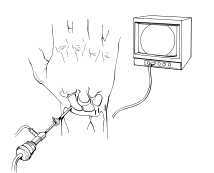
Figure 1
Schematic of arthroscope.
Copyright © American Society for Surgery of the Hand.
All content copied with permission from ASSH (www.assh.org). |